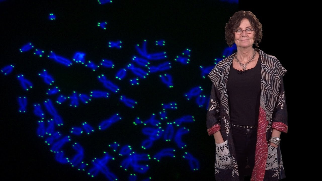
Protection and maintenance of mammalian telomeres
The continuity of life depends on the stable maintenance of genetic information. Eukaryotes have a special problem in this regard, stemming from the linear nature of their chromosomes. Chromosome ends pose two challenges: they require a specialized mechanism of duplication and they need to be protected from the cellular machinery that detects and repairs DNA breaks. We study telomeres, the elements that protect chromosome ends and ensure their complete replication.
Our lab focuses on the telomeres in mammalian cells, which are made up of long arrays of double-stranded TTAGGG repeats that end in a single-stranded 3’ overhang. This 3’ end is the substrate for telomerase, a reverse transcriptase that adds TTAGGG repeats to chromosome ends and thereby counteracts telomere attrition. Although telomerase is expressed in the germ line and certain stem cells, it is absent from most other human cells. As a consequence, many somatic cells undergo gradual telomere shortening and eventually arrest due to depletion of their telomere reserve. Cancer cells, on the other hand, often contain high levels of telomerase, which circumvents the replicative attrition of telomeric DNA and endows the cells with unlimited proliferative potential. The goal of our research is to understand how telomeres protect chromosome ends, how telomeres interact with and regulate telomerase, and what happens when telomere function is lost.
T-Loops | Relevant Publications
In collaboration with Jack Griffith at the University of North Carolina, Chapel Hill we found that telomeres can occur in a lariat conformation, referred to as the t-loop. T-loops are formed through the strand invasion of the 3’ telomeric overhang into the duplex part of the telomere. This structure is proposed to hide the telomere terminus from DNA repair enzymes that threaten the integrity of chromosome ends. Since the discovery of t-loops in mammals, they have been found in many other eukaryotes, including protozoa, plants, and some fungi.
Shelterin| Relevant Publications
We have identified a telomere-specific protein complex, called shelterin, that is crucial for the protection of telomeres from the DNA damage response and regulates telomere maintenance by telomerase. Shelterin is composed of six proteins: TRF1, TRF2, Rap1, TIN2, TPP1, and POT1. TRF1 and TRF2 bind to the duplex telomeric repeat array and anchor shelterin on telomeres. POT1 binds to single-stranded TTAGGG repeats and is recruited to telomeres through its interaction with TPP1. TPP1 in turns binds to TIN2, which interacts with both TRF1 and TRF2. Due to its specificity for the sequence and structure of the telomeric DNA, shelterin accumulates at the ends of human and mouse chromosomes but does not bind elsewhere in the genome. Thus, shelterin constitutes a unique marker of telomeres that allows cells to distinguish natural chromosome ends from sites of DNA damage. Our main approach to understanding the function of shelterin is to generate mouse cells from which individual shelterin proteins can be removed using inducible systems, e.g. Cre-mediated deletion.
Shelterin interacts with a large number of accessory factors that can be distinguished from the shelterin core components. The accessory factors are often transiently associated with telomeres, have functions elsewhere in the cell, and are less abundant at telomeres than shelterin. Many of the accessory factors are involved in DNA transactions (e.g. the Mre11 complex, Ku70/80, the Apollo nuclease, the WRN and BLM helicases), whereas others are protein modifying enzymes (e.g. tankyrase 1 and 2). The challenge is to understand how shelterin uses these factors to promote telomere protection while blocking their potential detrimental effects at telomeres.
Shelterin is rather diverged from the telomeric protein complexes studied in unicellular organisms. For instance, orthologs of TRF1 and TRF2 are not found at telomeres in S. cerevisiae and TIN2 has not been found outside metazoans. The most conserved component of shelterin is POT1, which has orthologs at telomeres in ciliates, plants, and some fungi. Yet, even POT1 evolves rapidly. A striking example of the rapid evolutionary changes at telomeres is found in mammals. Most mammals have just one POT1 gene and a single POT1 is recognized in chickens and Xenopus. In contrast, rodents have recently duplicated their POT1 gene, resulting in two distinct POT1 proteins (POT1a and POT1b) that have different functions at telomeres. This duplication suggests that rodents have evolved beyond other mammals, at least with regard to their shelterin complex.
Inhibition of DNA repair by shelterin | Relevant Publications
Telomeres are threatened by the two major DNA repair pathways that can act on DNA ends: non-homologous end-joining (NHEJ) and homology directed repair (HDR). NHEJ at telomeres is rampant when TRF2 is deleted from mouse cells, resulting in long trains of joined chromosomes. The remarkable frequency of NHEJ at dysfunctional telomeres in the TRF2 null setting now provides us with a unique tool to study this important repair process. TRF2 is also involved in repression of HDR. The N-terminal domain of TRF2 is required to repress resolution of the t-loop structure by enzymes involved in HDR. This reaction, termed t-loop HR, results in truncation of telomeres by removing the part of the telomeric DNA that constitutes the loop of the t-loop. A second HDR reaction can lead to inappropriate sequence exchanges between telomeres, and this process is repressed by both TRF2 and the NHEJ factor Ku70.
Repression of ATM and ATR by shelterin | Relevant Publications
DNA lesions can activate two transducing kinases, ATM and ATR, which enforce cell cycle arrest through phosphorylation of effector kinases and other targets, such as p53. When TRF2 is inhibited in human cells or deleted mouse cells, telomeres are recognized as sites of DNA damage and activate the canonical ATM kinase pathway. DNA damage response factors such as 53BP1 and MDC1 accumulate near telomeres and the histone H2AX becomes phosphorylated at chromosome ends. The ATM kinase becomes autophosphorylated on S1981 and its signaling activity results in a p53-dependent cell cycle arrest. These responses are entirely dependent on ATM. When TRF2 is deleted from ATM deficient cells, no DNA damage response is observed, indicating that the ATR kinase is not activated by telomeres lacking TRF2. Conversely, ATR but not ATM is activated when POT1 is removed from telomeres. Thus, telomeres use two distinct shelterin components to repress the two main DNA damage signaling pathways. These findings suggest how telomeres induce cell cycle arrest when they become critically shortened. Since the amount of shelterin at telomeres is dependent on telomere length, shortened telomeres are expected to carry diminishing amounts of TRF2 and POT1 which will eventually lead to insufficient repression of ATM and ATR.
Control of the telomere terminus by POT1 | Relevant Publications
The structure of the telomere terminus is regulated by POT1. When POT1 is inhibited, the 5’ end of the telomeres is altered from its normal precise ATC-5’ structure to a random position within the AATCCC sequence. In mouse cells, deletion of POT1b also affects the structure of the telomere terminus. POT1b deficient cells have unusually long 3’ overhang segments, in some cases more than 5 times longer than the wild type situation. Most likely, the long overhangs are due to excessive exonucleolytic attack on the 5’ ends of telomeres. The nature of the nuclease that threatens chromosome ends is not known.
Regulation of telomerase | Relevant Publications
In telomerase positive cells, telomeres are usually maintained at a stable length setting. This homeostasis is due to a negative feedback loop that controls the action of telomerase at individual chromosome ends. Shelterin is a key component of this control pathway, acting as a negative regulator of telomerase. As a telomere grows longer, it contains more shelterin, resulting in diminished activity of telomerase at that particular telomere end. Conversely, short telomeres have less shelterin and therefore exert less inhibition on telomerase. The inhibition of telomerase is largely dependent on the ability of shelterin to load POT1 on the single-stranded telomeric DNA. A mutant form of POT1 that loads on telomeres but lacks the ability to engage the 3’ overhang abrogates telomere length control even though all other components of shelterin are present.
